Tungiasis: Symptoms And Treatment
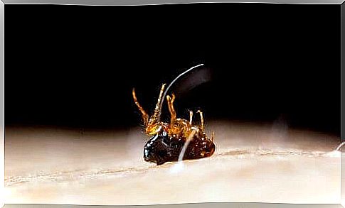
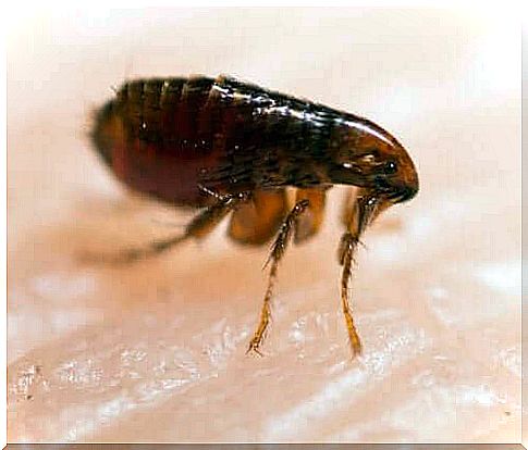
Tungiasis is an ectoparasitosis. In other words, it is a disease of parasites. It is caused by the sand flea, heavy penetrans. This is an insect smaller than 0.76 mm that attaches to the skin and produces an intense itching. It primarily attacks the feet.
The disease occurs in tropical areas and jungle areas of America, Africa and Asia. It has become more and more rare due to the development of society. The use of footwear and floors made of clinker and cement prevents the spread of it.
The flea, which is responsible for the development of tungiasis, has low host specificity. The term “host” refers to the body that allows the flea to survive.
In addition to humans, this condition can also affect poultry, dogs and pigs. The habitats where it is more common to find this flea consist of dry, sandy, shady and temperate soil. They also live in floors in sheds, houses and stables.
Symptoms of tungiasis
The disease causes various symptoms. It typically hits the feet. However, cases of infection of the legs, knees, thighs, hands, elbows and other parts of the body have been reported.
The lesions may be simple or numerous. They may itch, hurt or otherwise be completely asymptomatic. The phenomenon of flea infestation is asymptomatic. After 24 hours, however, the affected person may begin to see an itchy or reddish spot.
The place where the flea penetrated, the affected person can also see whitish nodules with a black dot in the middle, which corresponds to the stomach of the flea. They can also see eggs stuck to the skin close to the wound. When the flea dies, the wound has a black crust. This crust consists of clotted blood, among other things, and shrinks, leaving a scar on the skin.
Although tungiasis tends to disappear spontaneously after 4-6 weeks, it is common for it to recur. The patient usually also suffers from other concomitant infections, such as:
- Infection of the skin
- Abscesses
- Osteomyelitis
- Superficial varicose veins
- Inflammation of the lymphatic vessels
- In severe cases, blood poisoning and death
Some experts made a classification known as the Fortaleza classification to standardize the clinical description and facilitate the recognition of lesions as they develop.
Fortaleza classification
This classification consists of five stages, from penetration to shrinkage of the wound. There are less common clinical variations with dirty, pustular, ulcer-like and wart-like, such as foot warts and lesions.
In stage 1, the time that has elapsed since the intrusion is 30-120 minutes. During this stage, an erythematous macula appears at the site where the flea penetrated.
Stage 2 begins one to two days after infection. In this case, a hypochromic macula occurs with a black spot (which we have explained earlier, corresponding to the flea’s stomach), surrounded by an erythematous halo.
With stage 3, it corresponds to the time between 2 and 21 days after the intrusion. During that time, a whitish, painful spot with a diameter of 3 to 10 mm occurs with a black spot in the middle. The patient may also suffer from hyperkeratosis and yellowish pus. The patient can see the eggs coming from the flea.
When you move on to stage 4, it corresponds to the period between three and five weeks after the intrusion. At this stage, the flea dies. Then a halo of dead skin covered with crust will form around the original wound.
Eventually, in stage 5, which corresponds to about 6 weeks to several months after penetration, the wound disappears. In other words, it forms a small scar that disappears over time.
Treatment

The first step is to get the flea out while being careful not to ruin it. To that end, the medical professional usually enlarges the hole and presses on the edges to remove the parasite. They should do so under completely sterile circumstances.
They should also use a local antiseptic to prevent other infections and reduce the risk of complications. Likewise, they should provide a treatment for tetanus prophylaxis.